Uncategorized files
Appearance
Showing below up to 45 results in range #51 to #95.
-
LumbarPuntureforSA.jpg 351 × 135; 16 KB
-
Mapleson Circuits.jpeg 924 × 1,305; 298 KB
-
Measurements.png 495 × 295; 43 KB
-
Mesenteric Cysts.jpg 352 × 245; 31 KB
-
NMBcomparison.jpg 946 × 1,347; 224 KB
-
Needle misplacement.jpg 436 × 537; 63 KB
-
Numb and Number.pdf ; 23 KB
-
Orientation of the needle during puncture.jpg 435 × 163; 18 KB
-
Oxyhaemoglobin dissociation curve.png 439 × 413; 39 KB
-
PALS.pdf ; 150 KB
-
PALS Shaffner.jpg 517 × 667; 87 KB
-
Pedi-Crisis-new-logo.png 884 × 878; 88 KB
-
Pediatric lung isolation info sheet.jpg 2,481 × 3,509; 808 KB
-
Peds vs Adult Airway.jpg 547 × 789; 34 KB
-
Periop Insulin Pump Guidelines.pdf ; 246 KB
-
Posterior aspect of sacrum.jpg 287 × 284; 34 KB
-
Prone Chest Compression.jpg 988 × 511; 91 KB
-
PsoasCompartmentBlock.jpg 350 × 313; 25 KB
-
Puncture - orientation of the needle and reorientation after.jpg 410 × 252; 36 KB
-
Soft-tissue lateral neck radiograph.png 360 × 244; 79 KB
-
SpinalCord.jpg 307 × 432; 29 KB
-
SpinalFormula.jpg 350 × 44; 9 KB
-
SpinalNeedlesforSA.jpg 123 × 167; 6 KB
-
TYK102.png 305 × 71; 49 KB
-
TYK105.png 382 × 62; 52 KB
-
TYK99.png 374 × 53; 47 KB
-
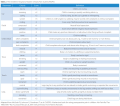
Table.png 1,013 × 787; 133 KB
-
Table 2.png 1,053 × 931; 169 KB
-
Test image.jpg 259 × 195; 8 KB
-
TymoteuszKajstura.jpg 1,913 × 2,889; 842 KB
-
US of sacro-coccygeal space.jpg 436 × 295; 20 KB
-
US use in caudal block.jpg 437 × 166; 20 KB
-
Ultrasound landmarks for TAP block.jpg 435 × 290; 19 KB
-
Ultrasound landmarks for iliinguinal-iliohypogastric.jpg 439 × 297; 19 KB
-
Ultrasound landmarks for rectus sheath block.jpg 438 × 290; 18 KB
-
Ultrasound probe position for iliinguinal-iliohypogastric.jpg 438 × 324; 23 KB
-
Ultrasound probe position for rectus sheath block.jpg 429 × 291; 20 KB
-
Ultrasound probe position for the transversus abdominis plane.jpg 435 × 325; 32 KB
-
Wfsahq-logo.png 236 × 68; 16 KB
-
Wilms Tumor Incision.jpg 351 × 286; 26 KB
-
Wilms Tumor Intraoperative.jpg 350 × 262; 28 KB
-
Wilms Tumor Pathology.jpg 349 × 262; 29 KB
-
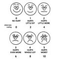
Wong-Baker Faces.jpg 296 × 297; 25 KB


